








IIT Gandhinagar Scientists Develop Breakthrough Plant Imaging Tool
GANDHINAGAR : Researchers from the Indian Institute of Technology Gandhinagar (IITGN) and the Regional Centre for Biotechnology (RCB), Faridabad, have developed a breakthrough imaging technology that...

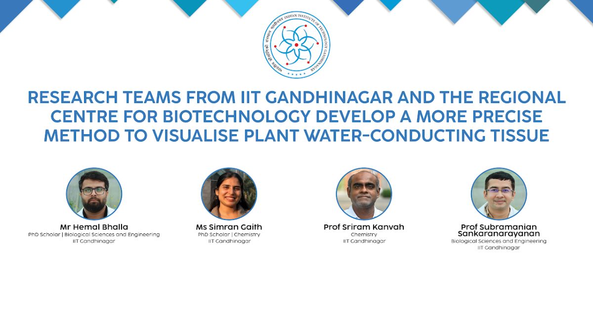
GANDHINAGAR : Researchers from the Indian Institute of Technology Gandhinagar (IITGN) and the Regional Centre for Biotechnology (RCB), Faridabad, have developed a breakthrough imaging technology that could transform plant science and accelerate the development of climate-resilient crops. Their newly designed fluorescent probes enable scientists to clearly visualise xylem, the plant tissue responsible for transporting water and minerals, with unprecedented precision.
The study, published in the journal Plant and Cell Physiology, introduces a faster, more sensitive and highly specific staining method that overcomes the limitations of conventional dyes used for decades in plant biology.
Xylem plays a critical role in helping plants survive drought, heat and other environmental stresses. However, existing dyes often stain multiple plant tissues simultaneously, making it difficult for researchers to distinguish xylem from surrounding tissues.
“The practical consequence is that a plant biologist looking at a stained section is often looking at xylem, phloem and cambium all lit up together, and has to reason about which glow belongs to which,” said Dr. Subramanian Sankaranarayanan, Assistant Professor in IITGN’s Department of Biological Sciences and Engineering.
The breakthrough came from an unexpected direction. Fluorescent molecules originally developed in 2023 for imaging mammalian cells were found to selectively identify plant xylem because of their sensitivity to different chemical environments.
“This crossover was leveraged by us,” said Prof. Sriram Kanvah, Professor in the Department of Chemistry at IITGN and the principal investigator of the project.
The research team tested four pyridinium-based fluorescent probes. Two of them, C1 and C3, produced remarkably sharp images of xylem while leaving surrounding tissues largely unstained. The probes were successfully evaluated in Arabidopsis thaliana, Nicotiana benthamiana, and Welsh onion, demonstrating their potential across different plant species.
The researchers also found that the new probes required only 25 micromolar concentration to produce clear images, compared with 375 micromolar needed for the widely used propidium iodide dye. This means scientists can achieve high-quality imaging using nearly 15 times less dye while reducing photobleaching and unwanted background staining.
The probes also performed exceptionally well in imaging damaged xylem vessels in mutant plants, offering researchers a powerful tool to study vascular defects and plant diseases.
“The project brought together molecular design, spectroscopy and plant biology to develop probes with properties tailored for biological imaging,” Prof. Kanvah said. “It was particularly exciting to see molecules originally developed for a different application perform so effectively in plant vascular tissues.”
Dr. Sankaranarayanan added that the research is aimed at improving scientific tools rather than answering a single biological question.
“By increasing the sensitivity and specificity of xylem staining, we hope these probes will make it easier to investigate plant development, vascular defects, and responses to environmental conditions,” he said.
The researchers believe the technology could eventually allow scientists to observe water-conducting tissues forming and responding to environmental stress in real time. Such advances could support the development of stronger, more resilient crops capable of withstanding the growing impacts of climate change.